What is Magnetic Resonance Imaging?
Magnetic Resonance Imaging (MRI) is a non-invasive medical imaging technique that uses strong magnetic fields and radio waves to create detailed images of the organs and tissues within the body.
Unlike X-rays or CT scans, MRI does not involve ionizing radiation, making it a safer option for many patients.
Definition of MRI
MRI, or Magnetic Resonance Imaging, is a diagnostic tool that utilizes the principles of nuclear magnetic resonance to generate images. By aligning the protons in the body’s hydrogen atoms with a strong magnetic field and then disrupting this alignment with radiofrequency pulses,
MRI captures the resulting signals to produce high-resolution images of internal structures. This technology is particularly effective for imaging soft tissues, making it invaluable in diagnosing conditions affecting the brain, spine, muscles, and joints.
History and Development of MRI Technology
The development of MRI technology began in the 1970s, building upon earlier research in nuclear magnetic resonance (NMR), which was primarily used in chemistry. In 1973, Dr. Raymond Damadian conducted the first full-body MRI scan, and in 1977, Dr. Richard Ernst and Dr.
Peter Mansfield made significant advancements that led to practical MRI imaging techniques. The first clinical MRI scans were performed shortly after, and by the 1980s, MRI had gained widespread acceptance in the medical community.
Since then, continuous improvements in technology have enhanced image quality and reduced scan times, making MRI a cornerstone of modern medical imaging.
How Does MRI Work?
MRI works by utilizing strong magnetic fields, radio waves, and advanced computer technology to generate detailed images of the body’s internal structures. The process relies on the principles of nuclear magnetic resonance to visualize tissues that are often difficult to see with other imaging techniques.
The Science Behind MRI
The science of MRI is based on the behavior of hydrogen protons, which are abundant in the human body, particularly in water and fat. When a patient is placed inside the MRI scanner, the strong magnetic field aligns these protons.
A radiofrequency pulse is then applied, temporarily disrupting this alignment. Once the pulse is turned off, the protons return to their original state, releasing energy in the process.
This emitted energy is detected by the MRI machine and converted into images through complex algorithms. The differences in relaxation times and the energy emitted by different tissues allow for the creation of high-contrast images, making it easier to distinguish between various structures.
MRI Machines and Technology
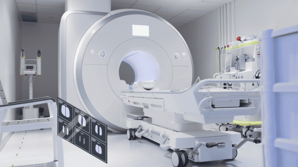
MRI machines consist of a large, cylindrical magnet, radiofrequency coils, and a computer system. The magnet creates a powerful magnetic field, while the radiofrequency coils send and receive radio waves. Modern MRI machines are equipped with advanced technology, such as high-field magnets (1.5 Tesla and above), which improve image quality and speed.
Some machines also feature open designs to accommodate patients who may feel claustrophobic. Additionally, innovations like functional MRI (fMRI) allow for the measurement of brain activity by detecting changes in blood flow, expanding the utility of MRI beyond standard imaging.
Types of MRI Scans
There are several types of MRI scans, each tailored for specific diagnostic purposes:
- Standard MRI: Provides detailed images of soft tissues, organs, and structures.
- Functional MRI (fMRI): Measures brain activity by detecting changes in blood flow, useful for neurological assessments.
- Magnetic Resonance Angiography (MRA): Focuses on blood vessels, helping to identify blockages or abnormalities.
- Diffusion MRI: Assesses the movement of water molecules in tissues, often used in brain imaging to detect strokes or tumors.
- Contrast MRI: Involves the use of a contrast agent to enhance the visibility of specific areas, often utilized in examining tumors or vascular conditions.
Each type of MRI scan serves a unique purpose, allowing healthcare providers to obtain critical information for diagnosis and treatment planning.
Benefits of Magnetic Resonance Imaging
Magnetic Resonance Imaging (MRI) provides a range of significant advantages that make it a vital diagnostic tool in contemporary medicine. Its capabilities allow for comprehensive evaluations of various medical conditions while emphasizing patient safety and comfort. Let’s explore these benefits in detail.
Non-Invasive Imaging
One of the standout features of MRI is its non-invasive nature. This imaging technique enables healthcare providers to obtain detailed images of the body’s internal structures without the need for any surgical procedures. Patients lie comfortably within the MRI machine, which does not require any cuts, injections, or other invasive measures.
This aspect is particularly beneficial for those who may be anxious about medical procedures or for children, as it reduces the overall stress associated with imaging. The non-invasive approach not only promotes patient comfort but also facilitates earlier diagnosis and management of conditions.
With MRI, physicians can assess a wide array of health issues—from joint disorders to neurological conditions—without resorting to more invasive diagnostic methods, which can carry higher risks.
Detailed Soft Tissue Visualization
MRI is uniquely suited for providing high-resolution images of soft tissues, making it invaluable for diagnosing a variety of medical conditions. While other imaging modalities, such as X-rays and CT scans, excel in visualizing bone structures, they often fall short in differentiating between various types of soft tissues.
MRI, on the other hand, offers superior contrast between different tissue types, allowing for clearer visualization of organs, muscles, cartilage, and even nerves.
This capability is essential in diagnosing conditions like tumors, where MRI can reveal not only the size and location of a mass but also its relationship to surrounding tissues.
Furthermore, MRI is particularly effective in identifying inflammatory diseases, ligament tears, and degenerative conditions, providing detailed information that aids in formulating accurate treatment plans. The ability to visualize soft tissues in multiple planes and sequences enhances diagnostic precision, ultimately leading to better patient outcomes.
Safety and No Radiation Exposure
A key advantage of MRI is its excellent safety profile, primarily due to the absence of ionizing radiation. Traditional imaging methods like X-rays and CT scans expose patients to radiation,
which can pose risks, especially with repeated exposures over time. In contrast, MRI uses strong magnetic fields and radio waves to produce images, eliminating the concerns associated with radiation exposure.
This safety factor is particularly crucial for specific patient populations, including children, pregnant women, and individuals requiring multiple follow-up scans.
Because MRI does not carry the same risks as radiation-based imaging, it can be performed repeatedly as needed for ongoing evaluation of a patient’s condition without increasing health risks.
Preparing for an MRI
Preparation is key to ensuring a smooth and successful MRI experience. Understanding what to expect and how to get ready can help alleviate anxiety and ensure accurate imaging results.
What to Expect Before the Scan
Before your MRI appointment, you will likely receive instructions from your healthcare provider or the imaging facility. It’s important to arrive on time to complete any necessary paperwork. You may be asked about your medical history, including any existing health conditions, allergies, and medications you are taking.
In some cases, you might need to change into a hospital gown and remove any metal objects, such as jewelry, watches, or hairpins, as these can interfere with the magnetic field. If you have a pacemaker, implantable devices, or other metal implants, inform the staff, as these may affect your eligibility for an MRI.
Preparing Physically and Mentally
While MRI scans are non-invasive, some patients may feel anxious about the procedure, especially if they are claustrophobic. To prepare mentally, consider discussing any concerns with your doctor or the MRI technologist beforehand. They can provide reassurance and explain the process to help alleviate anxiety.
Physically, you might be asked to refrain from eating or drinking for a few hours prior to your scan, particularly if a contrast agent will be used. Follow any specific guidelines provided by your healthcare team to ensure optimal imaging results.
What to Bring to Your Appointment
When heading to your MRI appointment, bring any relevant medical documents, including your doctor’s referral, insurance information, and identification.
If you have previous imaging studies (like X-rays or CT scans), consider bringing those along for comparison. Additionally, wear comfortable clothing that is free from metal, or be prepared to change into a gown provided by the facility.
The MRI Procedure
Understanding the MRI procedure can help demystify the experience and reduce any apprehensions you may have.
Step-by-Step Overview of the MRI Process
- Arrival and Check-In: Arrive at the facility, check in, and complete any necessary paperwork.
- Changing: If required, change into a gown and remove any metallic items.
- Positioning: The MRI technologist will help you onto the MRI table. You may be asked to lie still, and cushions or straps may be used to keep you comfortable and in position.
- Scanning: Once you are positioned, the table will slide into the MRI machine. The technologist will step outside the scanning room but will be able to see and communicate with you through an intercom.
- Imaging: During the scan, you’ll hear loud tapping or thumping noises as the machine operates. It’s important to remain still to ensure clear images.
- Completion: After the imaging is complete, the technologist will assist you in getting off the table, and you can resume normal activities unless otherwise instructed.
Duration of the Scan
The length of an MRI scan can vary depending on the area being examined and whether contrast is used. Generally, the scan can take anywhere from 15 to 90 minutes. For example, a brain MRI might take about 30 minutes, while a comprehensive scan of the abdomen may take longer. Your healthcare provider will give you an estimate of how long your specific scan will take.
Possible Sensations During the Scan
While undergoing an MRI, most patients experience minimal discomfort. However, some sensations are common, including:
- Noise: The MRI machine can be quite loud, with banging and thumping sounds during the imaging process. Earplugs or headphones are often provided to help reduce noise levels.
- Tight Spaces: If you have claustrophobia, the enclosed space of the MRI machine may be challenging. Open MRI machines are available at some facilities, providing a more spacious environment.
- Coolness: The MRI machine is often kept cool, so you may feel a bit chilly. A blanket can be offered for added comfort.
- Contrast Reactions: If a contrast agent is used, you might feel a brief warm sensation as it is injected, but this typically subsides quickly.
Understanding MRI Results
After your MRI, interpreting the results is a crucial step in your diagnostic journey.
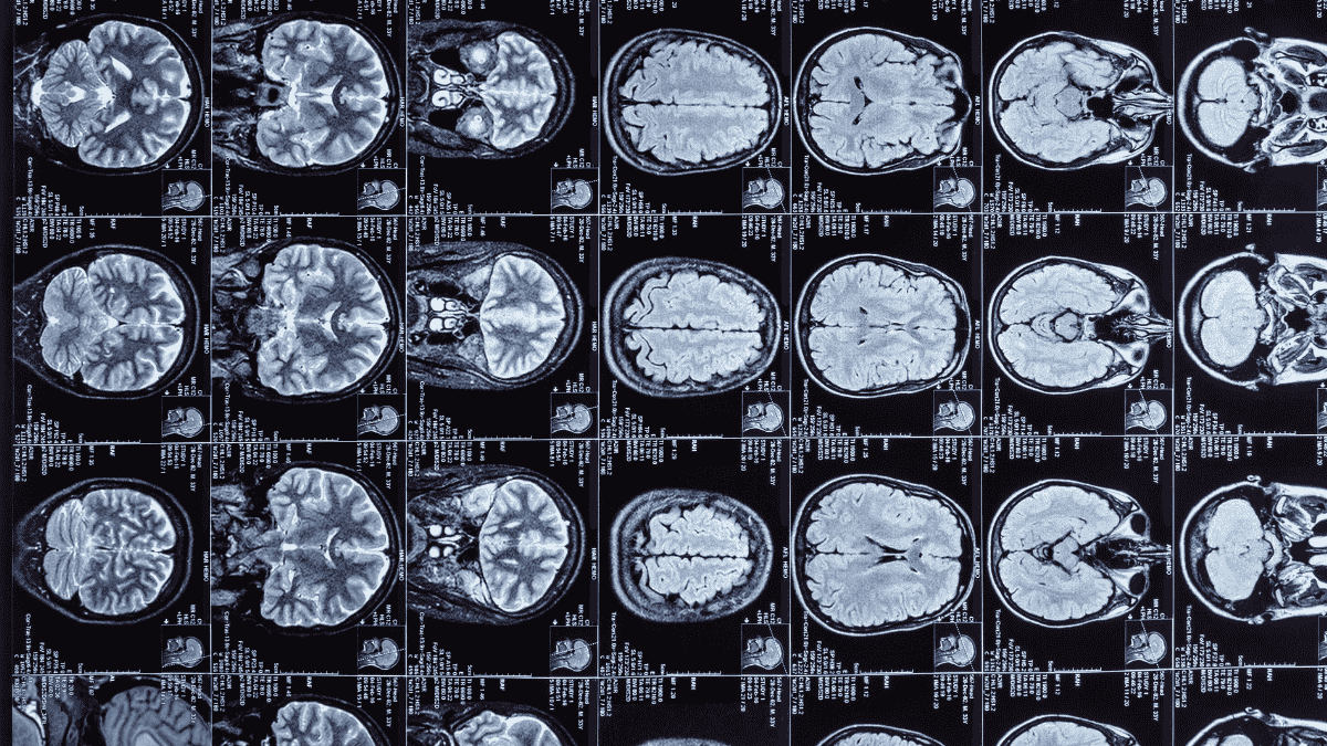
How MRI Images are Interpreted
Once the MRI scan is complete, a radiologist—an expert in medical imaging—will review the images. They will analyze the images for any abnormalities, such as tumors, inflammation, or structural issues. The radiologist will then compile a report detailing their findings, which will be sent to your referring physician.
It’s important to note that some results may take time to interpret fully, especially if additional imaging or assessments are required. Your doctor will discuss the results with you during a follow-up appointment.
Common Conditions Diagnosed with MRI
MRI is an effective diagnostic tool for a variety of conditions, including:
- Neurological Disorders: Tumors, multiple sclerosis, and stroke assessments.
- Joint and Muscle Issues: Torn ligaments, arthritis, and soft tissue injuries.
- Spinal Conditions: Herniated discs, spinal stenosis, and abnormalities.
- Cardiovascular Problems: Assessments of blood vessels and heart conditions through Magnetic Resonance Angiography (MRA).
These examples illustrate the broad applicability of MRI in diagnosing a wide range of health concerns.
Follow-Up and Next Steps
After your doctor receives the MRI results, they will discuss the findings with you and recommend any necessary follow-up actions. Depending on the results, your doctor may suggest further imaging, referrals to specialists, or specific treatment plans.
It’s essential to engage in this discussion and ask any questions you may have about your diagnosis, treatment options, and any additional tests that may be needed. Understanding the next steps is key to managing your health effectively.
Conclusion
Magnetic Resonance Imaging (MRI) stands out as a crucial tool in modern healthcare, offering numerous benefits that enhance diagnostic accuracy and patient safety.
Its non-invasive nature allows for detailed visualization of soft tissues, making it invaluable in identifying a wide range of medical conditions, from neurological disorders to musculoskeletal injuries.
Preparation for an MRI, both physically and mentally, can help alleviate anxiety and ensure a smooth experience. Understanding the MRI procedure itself—from what to expect before the scan to how results are interpreted—empowers patients to be active participants in their healthcare journey.
FAQs
Is MRI safe for everyone?
Yes, but certain conditions (like pacemakers) may require caution.
How does MRI compare to other imaging techniques?
MRI provides superior soft tissue contrast without radiation exposure.
What should I do if I’m claustrophobic?
Discuss your concerns with the staff; open MRI options may be available.
Do I need to prepare for the scan?
Yes, follow any specific guidelines provided by your healthcare team.
How long does an MRI take?
Typically between 15 to 90 minutes, depending on the area being scanned.
Will I feel pain during the MRI?
No, but you may experience some noise and a sense of tightness.